Biological roles

Containment and separationedit
The primary role of the lipid bilayer in biology is to separate aqueous compartments from their surroundings. Without some form of barrier delineating “self” from “non-self,” it is difficult to even define the concept of an organism or of life. This barrier takes the form of a lipid bilayer in all known life forms except for a few species of archaea that utilize a specially adapted lipid monolayer. It has even been proposed that the very first form of life may have been a simple lipid vesicle with virtually its sole biosynthetic capability being the production of more phospholipids. The partitioning ability of the lipid bilayer is based on the fact that hydrophilic molecules cannot easily cross the hydrophobic bilayer core, as discussed in Transport across the bilayer below. The nucleus, mitochondria and chloroplasts have two lipid bilayers, while other sub-cellular structures are surrounded by a single lipid bilayer (such as the plasma membrane, endoplasmic reticula, Golgi apparatus and lysosomes). See Organelle.
Prokaryotes have only one lipid bilayer - the cell membrane (also known as the plasma membrane). Many prokaryotes also have a cell wall, but the cell wall is composed of proteins or long chain carbohydrates, not lipids. In contrast, eukaryotes have a range of organelles including the nucleus, mitochondria, lysosomes and endoplasmic reticulum. All of these sub-cellular compartments are surrounded by one or more lipid bilayers and, together, typically comprise the majority of the bilayer area present in the cell. In liver hepatocytes for example, the plasma membrane accounts for only two percent of the total bilayer area of the cell, whereas the endoplasmic reticulum contains more than fifty percent and the mitochondria a further thirty percent.
Signalingedit
Probably the most familiar form of cellular signaling is synaptic transmission, whereby a nerve impulse that has reached the end of one neuron is conveyed to an adjacent neuron via the release of neurotransmitters. This transmission is made possible by the action of synaptic vesicles loaded with the neurotransmitters to be released. These vesicles fuse with the cell membrane at the pre-synaptic terminal and release its contents to the exterior of the cell. The contents then diffuse across the synapse to the post-synaptic terminal.
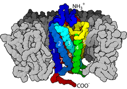
Lipid bilayers are also involved in signal transduction through their role as the home of integral membrane proteins. This is an extremely broad and important class of biomolecule. It is estimated that up to a third of the human proteome are membrane proteins. Some of these proteins are linked to the exterior of the cell membrane. An example of this is the CD59 protein, which identifies cells as “self” and thus inhibits their destruction by the immune system. The HIV virus evades the immune system in part by grafting these proteins from the host membrane onto its own surface. Alternatively, some membrane proteins penetrate all the way through the bilayer and serve to relay individual signal events from the outside to the inside of the cell. The most common class of this type of protein is the G protein-coupled receptor (GPCR). GPCRs are responsible for much of the cell's ability to sense its surroundings and, because of this important role, approximately 40% of all modern drugs are targeted at GPCRs.
In addition to protein- and solution-mediated processes, it is also possible for lipid bilayers to participate directly in signaling. A classic example of this is phosphatidylserine-triggered phagocytosis. Normally, phosphatidylserine is asymmetrically distributed in the cell membrane and is present only on the interior side. During programmed cell death a protein called a scramblase equilibrates this distribution, displaying phosphatidylserine on the extracellular bilayer face. The presence of phosphatidylserine then triggers phagocytosis to remove the dead or dying cell.
Comments
Post a Comment