Characterization methods

The lipid bilayer is a very difficult structure to study because it is so thin and fragile. In spite of these limitations dozens of techniques have been developed over the last seventy years to allow investigations of its structure and function.
Electrical measurementsedit
Electrical measurements are a straightforward way to characterize an important function of a bilayer: its ability to segregate and prevent the flow of ions in solution. By applying a voltage across the bilayer and measuring the resulting current, the resistance of the bilayer is determined. This resistance is typically quite high (108 Ohm-cm2 or more) since the hydrophobic core is impermeable to charged species. The presence of even a few nanometer-scale holes results in a dramatic increase in current. The sensitivity of this system is such that even the activity of single ion channels can be resolved.
Fluorescence microscopyedit
Electrical measurements do not provide an actual picture like imaging with a microscope can. Lipid bilayers cannot be seen in a traditional microscope because they are too thin. In order to see bilayers, researchers often use fluorescence microscopy. A sample is excited with one wavelength of light and observed in a different wavelength, so that only fluorescent molecules with a matching excitation and emission profile will be seen. Natural lipid bilayers are not fluorescent, so a dye is used that attaches to the desired molecules in the bilayer. Resolution is usually limited to a few hundred nanometers, much smaller than a typical cell but much larger than the thickness of a lipid bilayer.
Electron microscopyedit
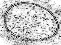
Electron microscopy offers a higher resolution image. In an electron microscope, a beam of focused electrons interacts with the sample rather than a beam of light as in traditional microscopy. In conjunction with rapid freezing techniques, electron microscopy has also been used to study the mechanisms of inter- and intracellular transport, for instance in demonstrating that exocytotic vesicles are the means of chemical release at synapses.
Nuclear magnetic resonance spectroscopyedit
31P-NMR(nuclear magnetic resonance) spectroscopy is widely used for studies of phospholipid bilayers and biological membranes in native conditions. The analysis of 31P-NMR spectra of lipids could provide a wide range of information about lipid bilayer packing, phase transitions (gel phase, physiological liquid crystal phase, ripple phases, non bilayer phases), lipid head group orientation/dynamics, and elastic properties of pure lipid bilayer and as a result of binding of proteins and other biomolecules.
Atomic force microscopyedit
A new method to study lipid bilayers is Atomic force microscopy (AFM). Rather than using a beam of light or particles, a very small sharpened tip scans the surface by making physical contact with the bilayer and moving across it, like a record player needle. AFM is a promising technique because it has the potential to image with nanometer resolution at room temperature and even under water or physiological buffer, conditions necessary for natural bilayer behavior. Utilizing this capability, AFM has been used to examine dynamic bilayer behavior including the formation of transmembrane pores (holes) and phase transitions in supported bilayers. Another advantage is that AFM does not require fluorescent or isotopic labeling of the lipids, since the probe tip interacts mechanically with the bilayer surface. Because of this, the same scan can image both lipids and associated proteins, sometimes even with single-molecule resolution. AFM can also probe the mechanical nature of lipid bilayers.
Dual polarisation interferometryedit
Lipid bilayers exhibit high levels of birefringence where the refractive index in the plane of the bilayer differs from that perpendicular by as much as 0.1 refractive index units. This has been used to characterise the degree of order and disruption in bilayers using dual polarisation interferometry to understand mechanisms of protein interaction.
Quantum chemical calculationsedit
Lipid bilayers are complicated molecular systems with many degrees of freedom. Thus, atomistic simulation of membrane and in particular ab initio calculations of its properties is difficult and computationally expensive. Quantum chemical calculations has recently been successfully performed to estimate dipole and quadrupole moments of lipid membranes.
Comments
Post a Comment